
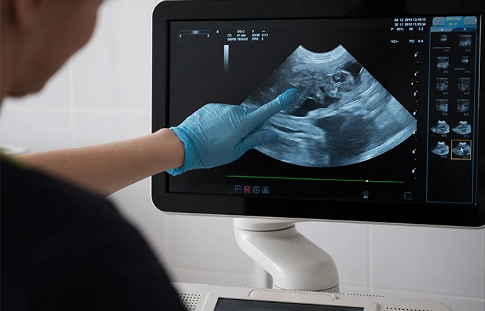
Diagnostic Sonography In Nagpur For Focused Imaging with Dr.Ritesh Nawkhare
Diagnostic Sonography In Nagpur offers real-time visual assessment of selected organs, soft tissues, blood vessels, muscles, and other accessible anatomical structures through high-frequency sound waves. The technique can provide dynamic information during an examination and may help healthcare professionals investigate unexplained symptoms, examine structural findings, assess selected circulation patterns, and monitor specific conditions. Since ultrasound does not use ionising radiation, it can be considered for a range of clinical situations when recommended by a qualified healthcare professional. At Neurosys Multispeciality Centre In Nagpur, sonography can be incorporated into a broader diagnostic evaluation based on the patient’s symptoms, medical history, examination findings, previous investigations, and specific clinical questions. Under the guidance of Dr. Ritesh Nawkhare, the diagnostic approach can be tailored to the patient’s individual requirements. Rather than treating ultrasound as a single type of scan, the examination can be selected according to the anatomical region and information that needs to be assessed.
Understanding Diagnostic Sonography
Diagnostic sonography uses a transducer to transmit high-frequency sound waves into the body and receive the returning echoes. Different tissues interact with sound waves in different ways, allowing the ultrasound system to generate images on a monitor. The examination can provide information about the appearance, location, size, and selected characteristics of accessible structures.
▪ Real-time visualisation of selected tissues
▪ Assessment of organ and soft-tissue structures
▪ Evaluation of selected vascular flow patterns
▪ Dynamic examination of certain musculoskeletal structures
▪ Pregnancy and fetal assessment when clinically indicated
▪ Support for selected image-guided procedures
The information obtained depends on the type of sonography performed and the structure being examined.
Advanced Sonography For Focused Assessment
Modern sonography can be tailored to address specific clinical questions rather than providing only a broad anatomical overview. The imaging approach may vary according to the symptoms, body region, suspected condition, previous medical findings, and the type of information required by the healthcare professional. Real-time ultrasound imaging allows selected organs, tissues, and structures to be examined while the examination is being performed. In some cases, additional techniques such as Doppler assessment or dynamic imaging may provide further information. At Neurosys Multispeciality Centre In Nagpur, sonography may form part of a broader diagnostic evaluation. The findings can be considered alongside the patient’s medical history, physical examination, laboratory investigations, previous imaging reports, and other relevant clinical information. This approach helps the healthcare team focus the examination on the specific concern and determine whether additional assessment or follow-up may be appropriate.
Conventional Ultrasound Assessment
Conventional ultrasound provides real-time images of selected internal organs, soft tissues, and anatomical structures using high-frequency sound waves. It may be recommended to investigate symptoms, evaluate a suspected abnormality, assess a previously identified finding, or monitor changes over time. The examination can provide useful information about the size, shape, position, texture, and appearance of accessible structures. Depending on the area being examined, ultrasound may help identify findings such as fluid-filled areas, cystic changes, stones, swelling, tissue irregularities, or other structural abnormalities.
▪ Assessment of selected organs and soft tissues
▪ Evaluation of symptoms such as pain, swelling, or discomfort
▪ Review of previously identified structural findings
▪ Monitoring of selected conditions over time
▪ Real-time imaging during the examination
The exact scope of the examination depends on the clinical requirement. Your healthcare professional can explain which structures need to be assessed and what information the ultrasound is intended to provide.
Dynamic Ultrasound Evaluation
One of the useful features of ultrasound is its ability to display structures in real time. This allows certain tissues to be observed not only at rest but also during specific movements when clinically appropriate. Dynamic imaging can therefore provide additional information in selected musculoskeletal and superficial soft-tissue evaluations. Muscles, tendons, ligaments, joints, and other accessible structures may behave differently during movement compared with a static position. Observing these changes can help the healthcare professional assess the relationship between movement and a particular symptom or finding.
▪ Real-time observation of selected anatomical structures
▪ Assessment of muscles and tendons during movement
▪ Evaluation of selected joints and superficial tissues
▪ Comparison of structures during different positions
▪ Additional information when static imaging alone may not answer the clinical question
Dynamic ultrasound is not required for every examination. It is selected when movement-based assessment may add clinically useful information.
Doppler-Based Assessment
Doppler techniques extend conventional ultrasound by providing information about blood movement through selected vessels. Depending on the examination, Doppler imaging may help assess the direction and characteristics of blood flow and identify changes that may require further clinical evaluation. Doppler assessment can be used in several clinical settings, including selected vascular examinations and pregnancy-related imaging when blood-flow information is clinically indicated. The specific Doppler technique depends on the vessel, organ, or region being assessed.
▪ Evaluation of blood-flow patterns
▪ Assessment of selected blood vessels
▪ Identification of changes requiring further evaluation
▪ Additional information alongside structural ultrasound findings
▪ Pregnancy-related Doppler assessment when clinically recommended
Doppler findings are interpreted together with the clinical history and other examination findings rather than being considered in isolation.
Abdominal Sonography
Abdominal sonography provides real-time imaging of several organs and structures within the abdominal cavity. It may be recommended when symptoms, physical examination findings, laboratory results, or previous investigations suggest the need for further assessment. Depending on the clinical question, the examination may include evaluation of the liver, gallbladder, bile ducts, pancreas, spleen, kidneys, urinary bladder, and other accessible abdominal structures. Abdominal ultrasound can help assess the appearance and structure of these organs and may identify findings such as stones, cystic areas, fluid collections, enlargement, swelling, or other structural changes. It may also be used as part of follow-up when a previous examination has identified a finding that requires monitoring.
Liver And Gallbladder Assessment
Ultrasound evaluates the liver and gallbladder, helping identify structural changes, gallstones, bile duct abnormalities, and other findings related to abdominal symptoms.
1.Kidney And Urinary System Imaging
Abdominal sonography examines the kidneys and urinary bladder. It may help assess suspected stones, urinary symptoms, structural changes, and bladder-related concerns.
2.Pancreas And Spleen Evaluation
Ultrasound can assess the visible portions of the pancreas and spleen, with findings interpreted alongside symptoms, clinical examination, and other investigations.
When May Diagnostic Sonography Be Recommended?
Your doctor may recommend sonography when symptoms, examination findings, or previous investigations indicate that ultrasound could provide useful additional information.
It may be considered for:
▪ Abdominal or pelvic symptoms
▪ Localised swelling or palpable findings
▪ Selected soft-tissue concerns
▪ Musculoskeletal symptoms
▪ Pregnancy-related assessment
▪ Selected vascular concerns
▪ Monitoring of previously identified findings
The appropriate investigation varies between patients. A healthcare professional should determine whether ultrasound is suitable for your particular situation.
Benefits Of Diagnostic Sonography
Diagnostic sonography offers several practical advantages when ultrasound imaging is clinically appropriate:
▪ Non-invasive examination without surgical intervention
▪ Uses sound waves rather than ionising radiation
▪ Provides real-time images of selected organs and tissues
▪ Can assess certain structures during movement
▪ Doppler techniques can provide blood-flow information
▪ Useful for abdominal, pelvic, pregnancy, vascular, and musculoskeletal assessment
▪ Generally well tolerated and suitable for many age groups
▪ Can support diagnosis, monitoring, and follow-up of selected conditions
▪ May complement physical examination, laboratory tests, and other imaging studies.
Sonography As Part Of A Broader Diagnostic Approach
Ultrasound can provide valuable information, but it is not designed to replace every other diagnostic test. Some conditions or anatomical areas may require a different imaging method or additional investigation.
Depending on the clinical question, your healthcare provider may consider:
▪ MRI
▪ CT
▪ X-ray
▪ Blood investigations
▪ Other specialised diagnostic tests
The choice depends on the information needed to understand the patient’s condition.
Recovery After Diagnostic Sonography
Diagnostic sonography is generally a non-invasive examination, so most people can return to their usual activities immediately after the scan.
▪ No surgical recovery period is usually required
▪ Normal daily activities can generally be resumed after the examination
▪ You may eat and drink normally once any required preparation restrictions are over
▪ If contrast, medication, or an ultrasound-guided procedure is involved, additional instructions may apply
▪ Your healthcare professional will explain any follow-up needed based on the findings
Why Choose Neurosys Multispeciality Centre In Nagpur?
At Neurosys Multispeciality Centre In Nagpur, diagnostic sonography can be considered within a wider clinical assessment rather than as an isolated investigation. Under the guidance of Dr. Ritesh Nawkhare, the evaluation can take into account the patient’s symptoms, clinical history, examination findings, and available diagnostic records. The approach focuses on obtaining information that is relevant to the patient’s clinical concern.
▪ Individualised diagnostic assessment
▪ Selection of imaging according to clinical requirements
▪ Real-time ultrasound evaluation
▪ Doppler assessment when indicated
▪ Pregnancy and fetal imaging when clinically required
▪ Musculoskeletal and superficial soft-tissue assessment
▪ Review of previous imaging when relevant
▪ Clinical correlation of imaging findings
Frequently Asked Questions
Book a Consultation for Diagnostic sonography in Nagpur
The appropriate sonography examination depends on the symptoms being investigated, the anatomical area involved, medical history, previous findings, and the diagnostic information required. Discussing these factors with a healthcare professional can help determine whether ultrasound is appropriate and what type of examination may be suitable. At Neurosys Multispeciality Centre In Nagpur, patients can discuss their diagnostic requirements with the healthcare team under the guidance of Dr. Ritesh Nawkhare. Previous ultrasound reports, imaging studies, prescriptions, and relevant medical records can be brought to the consultation when available.
Following the recommended preparation instructions before the examination can also help support an effective imaging assessment.