Vascular ultrasound
Vascular ultrasound is a non-invasive imaging technique used to evaluate the circulatory system, including arteries and veins. This diagnostic tool is essential for detecting blockages, clots, and other abnormalities that can affect blood flow. By using high-frequency sound waves, vascular ultrasound provides detailed images of blood vessels, helping healthcare professionals diagnose and manage various vascular conditions.
The Importance of Vascular Ultrasound in Modern Medicine
Vascular ultrasound plays a crucial role in modern medicine. It allows for the early detection of vascular diseases, which can prevent severe complications such as stroke, heart attack, and deep vein thrombosis. The procedure is safe, painless, and does not involve radiation, making it a preferred choice for both patients and healthcare providers.
Types of Vascular Ultrasound
- Doppler Ultrasound: This type of ultrasound measures the speed and direction of blood flow. It is particularly useful for detecting blockages and assessing the severity of vascular diseases.
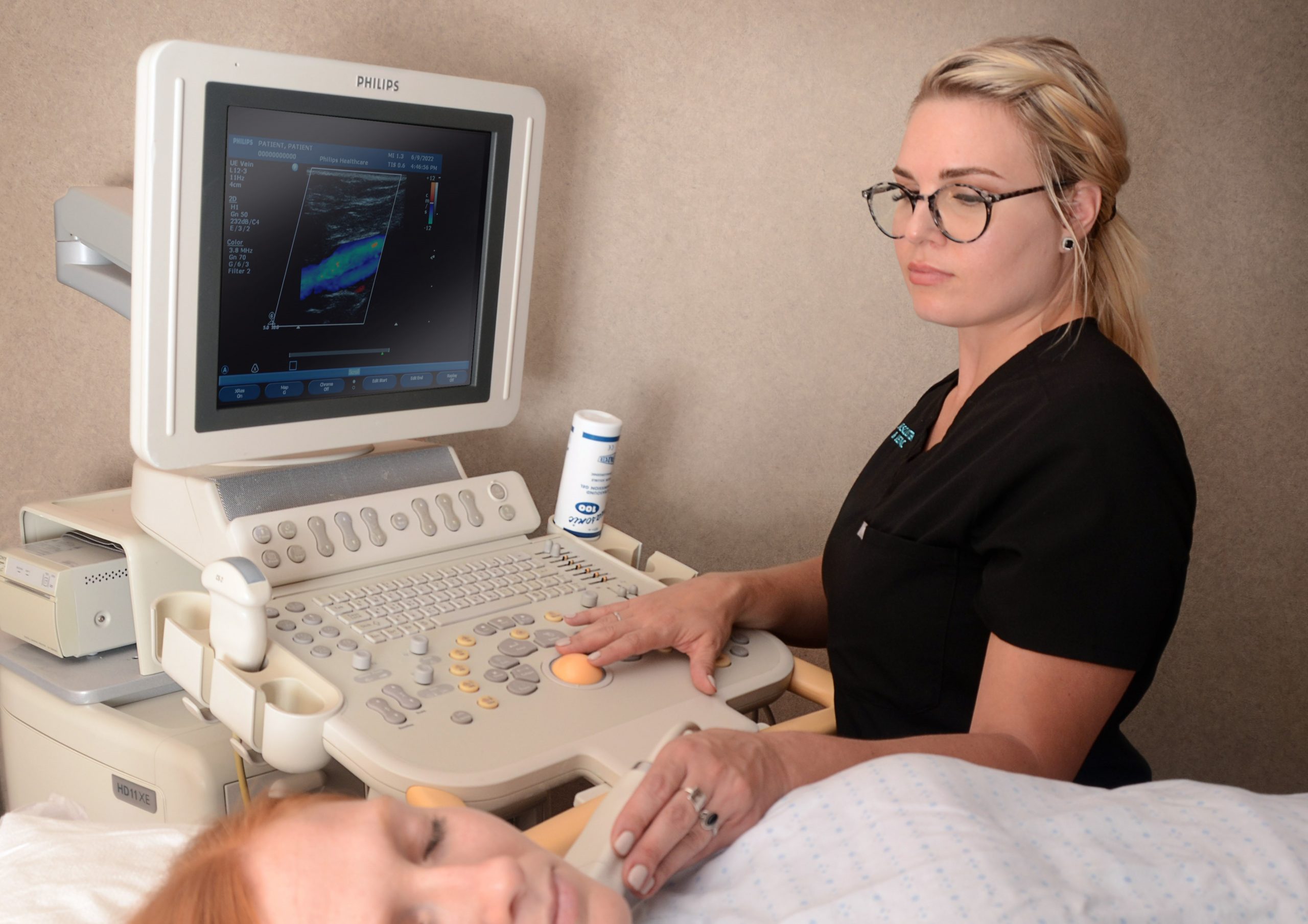
- Color Doppler Ultrasound: This technique adds color to the Doppler ultrasound images, making it easier to visualize blood flow and identify abnormalities.
- Duplex Ultrasound: Combining traditional ultrasound and Doppler ultrasound, duplex ultrasound provides both images and blood flow information, offering a comprehensive view of the vascular system
Applications of Vascular Ultrasound
Diagnosing Arterial Diseases
Vascular ultrasound is widely used to diagnose arterial diseases such as peripheral artery disease (PAD) and carotid artery disease. It helps in identifying plaque buildup, stenosis (narrowing of arteries), and aneurysms (abnormal bulging of artery walls).
Evaluating Venous Disorders
Venous disorders like deep vein thrombosis (DVT) and varicose veins can be effectively evaluated using vascular ultrasound. The procedure helps in detecting blood clots, assessing valve function, and guiding treatment plans.
Monitoring Vascular Health
Patients with chronic conditions such as diabetes and hypertension are at a higher risk of developing vascular complications. Regular vascular ultrasound exams can help monitor vascular health and detect issues early, preventing severe outcomes.
Pre- and Post-Surgical Assessments
Vascular ultrasound is often used before and after surgical procedures to assess the condition of blood vessels. It helps in planning surgeries, such as bypass grafts, and in monitoring the success of interventions like angioplasty
Advantages of Vascular Ultrasound
Non-Invasive and Painless
One of the most significant advantages of vascular ultrasound is that it is non-invasive and painless. Unlike other imaging techniques that may require incisions or the use of contrast dyes, vascular ultrasound is performed externally, making it a comfortable experience for patients.
No Radiation Exposure
Vascular ultrasound does not involve the use of ionizing radiation, making it a safer option compared to X-rays and CT scans. This is particularly beneficial for pregnant women and patients who require frequent imaging.
Real-Time Imaging
Vascular ultrasound provides real-time images, allowing healthcare providers to observe blood flow and vessel structures dynamically. This immediate feedback is invaluable during diagnostic procedures and interventions.
Cost-Effective
Compared to other imaging modalities, vascular ultrasound is relatively cost-effective. It provides detailed information without the high costs associated with MRI or CT scans, making it accessible to a broader population.
Preparing for a Vascular Ultrasound
What to Expect Before the Procedure
Before undergoing a vascular ultrasound, patients may be advised to follow specific instructions, such as fasting or avoiding certain medications. It is essential to inform the healthcare provider about any existing medical conditions or allergies.
During the Procedure
During the procedure, the patient will lie down on an examination table. A gel is applied to the skin to facilitate the transmission of sound waves. The ultrasound technician will then move a transducer over the area of interest, capturing images and blood flow data.
After the Procedure
Vascular ultrasound provides real-time images, allowing healthcare providers to observe blood flow and vessel structures dynamically. This immediate feedback is invaluable during diagnostic procedures and interventions.
Advances in Vascular Ultrasound Technology
3D and 4D Ultrasound
Recent advancements in ultrasound technology have introduced 3D and 4D imaging, providing even more detailed views of vascular structures. These techniques offer enhanced visualization, particularly useful in complex cases and surgical planning.
Contrast-Enhanced Ultrasound
Contrast-enhanced ultrasound involves the use of microbubble contrast agents to improve the clarity of ultrasound images. This technique is particularly beneficial in assessing blood flow and detecting small lesions that may be missed in standard ultrasound exams.
Portable Ultrasound Devices
The development of portable ultrasound devices has revolutionized vascular imaging, allowing for point-of-care diagnostics. These devices are especially useful in emergency settings, rural areas, and during surgical procedures.Contact Us
Conclusion
Vascular ultrasound is an indispensable tool in modern medicine, offering a safe, non-invasive, and cost-effective means of evaluating the circulatory system. Its applications range from diagnosing arterial and venous diseases to monitoring vascular health and guiding surgical interventions. With ongoing advancements in technology, vascular ultrasound continues to evolve, enhancing its diagnostic capabilities and expanding its reach. As healthcare providers and patients alike recognize the value of early detection and preventive care, vascular ultrasound will remain a cornerstone in the management of vascular diseases..Schedule your Consultation with Dr. Ritesh Nawkhare