
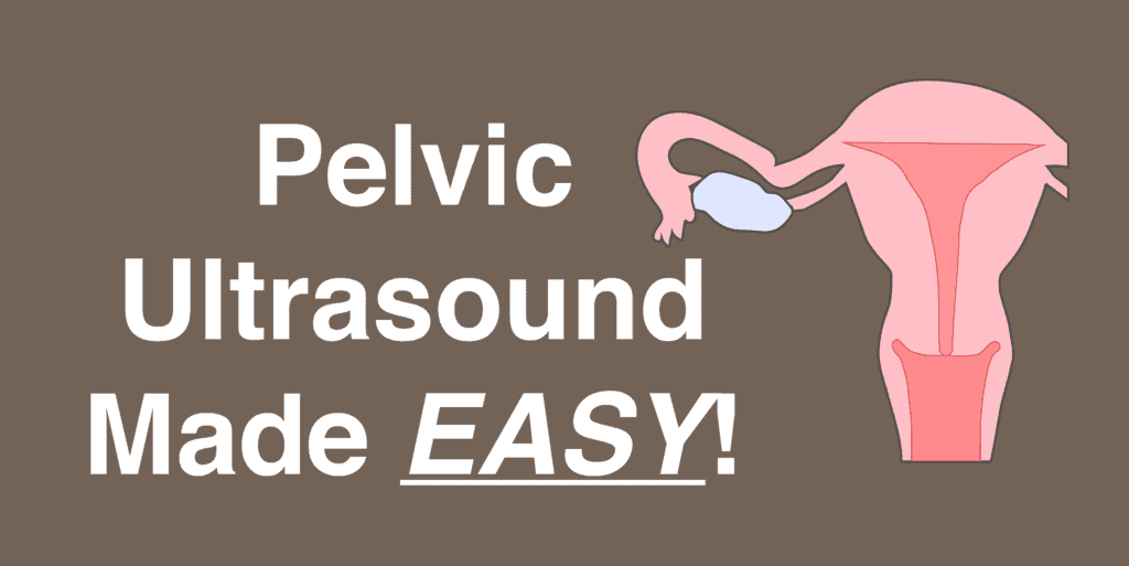
Pelvic Ultrasound In Nagpur | Detailed Imaging For Pelvic Health With Dr.Ritesh Nawkhare
Pelvic Ultrasound In Nagpur helps doctors examine the organs and structures within the lower abdomen and pelvic region. The scan can produce detailed images of the uterus, ovaries, cervix, urinary bladder, and nearby tissues. Doctors may recommend this examination when you have pelvic pain, unusual vaginal bleeding, menstrual changes, fertility concerns, or other symptoms that need further evaluation. At NNS Hospital, the medical team plans the ultrasound according to your symptoms, medical history, and clinical needs. The imaging approach can vary from patient to patient. A transabdominal ultrasound provides a broader view of the pelvic region. A transvaginal ultrasound can provide closer images of the uterus, uterine lining, ovaries, and other pelvic structures when clinically appropriate. The scan does more than create images. It helps doctors look for structural changes that may explain your symptoms. The findings can support further evaluation and help guide the next steps in your care. Depending on your symptoms and clinical requirements, doctors may also use Doppler imaging to assess blood flow through selected pelvic structures.
Understanding Your Pelvic Ultrasound In Nagpur
A pelvic ultrasound uses sound waves to create images of organs and structures within the pelvic region.
During the examination, your doctor may assess:
▪ The size, position, and appearance of the uterus
▪ The thickness and appearance of the uterine lining
▪ The size and structure of the ovaries
▪ The cervix and surrounding pelvic structures
▪ The urinary bladder
▪ Certain pelvic masses, cysts, or fluid-filled areas
▪ Blood flow in selected structures when Doppler imaging is required
The ultrasound findings can help your doctor understand possible causes of your symptoms. They can also support decisions about further testing or treatment. Depending on the findings, your doctor may recommend monitoring, additional imaging, laboratory tests, or other investigations. The next step will depend on your symptoms, medical history, and overall clinical assessment.
When Your Doctor May Recommend This Scan ?
Your doctor may advise a pelvic ultrasound for:
▪ Ongoing pain in the lower abdomen or pelvic region
▪ Heavy, prolonged, or irregular menstrual bleeding
▪ Unexplained bleeding between menstrual periods
▪ Pain or discomfort during menstruation
▪ Suspected ovarian cysts or other ovarian changes
▪ Possible uterine fibroids
▪ A feeling of pressure, heaviness, or fullness in the pelvis
▪ Unexplained swelling or a lump in the pelvic area
▪ Fertility-related assessment
▪ Monitoring of a previously diagnosed pelvic condition
▪ Follow-up after treatment or a previous abnormal finding
The reason for the ultrasound helps your doctor decide which structures need closer assessment.
Looking For The Cause Of Pelvic Pain
Pelvic pain can develop for many reasons. Finding the underlying cause helps your doctor plan the right evaluation. A pelvic ultrasound allows doctors to examine the pelvic organs and identify visible structural changes. The scan may help detect ovarian cysts, uterine fibroids, pelvic masses, fluid collections, or other abnormalities. Ultrasound findings can provide useful information, but imaging alone does not explain every cause of pelvic pain. Your doctor will also consider how long the pain has lasted and whether it occurs at specific times. They may ask about menstrual changes, bleeding, urinary symptoms, digestive symptoms, or other concerns. Your medical history and physical examination also help guide the assessment. If you experience sudden, severe, or rapidly worsening pelvic pain, seek medical attention promptly. Your doctor can assess your symptoms and decide whether you need an urgent examination or further investigations.
Assessing Changes In Menstrual Bleeding
Changes in your usual menstrual pattern may need further evaluation. A pelvic ultrasound helps doctors examine the uterus and ovaries when periods become unusually heavy, prolonged, painful, or irregular.
▪ The size, position, and appearance of the uterus
▪ The thickness and appearance of the uterine lining
▪ Fibroids, polyps, or other structural changes
▪ The size, shape, and appearance of the ovaries
▪ Certain cysts, masses, or other abnormal findings
▪ Fluid or other changes within the pelvic region
The ultrasound findings can help your doctor understand possible reasons for menstrual changes. However, the scan forms only one part of the overall assessment. Your doctor may also consider your menstrual history, symptoms, age, medical history, and examination findings.
Checking Ovarian Health During Pelvic Ultrasound In Nagpur
The ovaries can change in size or appearance for several reasons.
A pelvic ultrasound may help identify:
▪ Simple or complex ovarian cysts
▪ Enlarged ovaries
▪ Certain ovarian masses
▪ Changes in ovarian structure
▪ Findings that may require monitoring or further evaluation
An abnormal ultrasound finding does not always mean that you have a serious condition. Some ovarian changes may occur for benign or temporary reasons. Your doctor will interpret the ultrasound findings along with your age, symptoms, menstrual history, medical history, and examination results. If the scan shows a finding that needs further assessment, your doctor may recommend follow-up imaging or additional tests. This approach helps the medical team understand the finding more clearly and plan appropriate care based on your individual needs.
Examining The Uterus And Its Lining During Pelvic Ultrasound In Nagpur
The uterus plays an important role in menstrual and reproductive health.
Your doctor may recommend an ultrasound to investigate possible:
▪ Uterine fibroids
▪ Changes in the thickness or appearance of the uterine lining
▪ Structural abnormalities of the uterus
▪ Pelvic masses or other visible growths
▪ Fluid collections or other unusual findings
▪ Changes that require further monitoring
Ultrasound findings can provide useful information about the uterus and endometrium. However, not every abnormal finding indicates a serious condition. Your doctor will consider the scan results along with your symptoms, menstrual history, age, and medical history.
Role Of Ultrasound In Fertility Assessment
Pelvic ultrasound can support fertility evaluation by helping doctors examine the uterus and ovaries.
Depending on your clinical needs, the examination may assess:
▪ The size, position, and structure of the uterus
▪ The thickness and development of the endometrium
▪ The size and appearance of the ovaries
▪ The number, size, and growth of ovarian follicles
▪ Other pelvic findings that may be relevant to fertility
Ultrasound can provide valuable information, but fertility assessment usually involves more than one investigation. Your doctor may consider your menstrual history, symptoms, reproductive history, age, and other relevant factors. Based on your individual circumstances, the doctor can recommend the appropriate tests and explain what each result means for your evaluation.
Choosing The Right Ultrasound Approach
The type of pelvic ultrasound depends on your symptoms, medical history, and the structures that need closer assessment. Your doctor will choose the imaging approach that can provide the most useful information for your clinical needs.
▪ Transabdominal Ultrasound: The sonographer moves the ultrasound probe over the lower abdomen to create a broader view of the pelvic organs. You may need a comfortably full bladder for this examination.
▪ Transvaginal Ultrasound: The sonographer uses a specialised probe to obtain closer images of the uterus, endometrium, ovaries, and nearby pelvic structures. Your doctor may recommend this method when it can provide more detailed information and is clinically appropriate.
▪ Doppler Ultrasound: Doppler imaging can be added to a pelvic ultrasound when your doctor needs to assess blood flow. It can provide additional information about circulation within selected pelvic structures.
Your doctor may use one method or combine different ultrasound techniques. The choice depends on the reason for the examination and the information needed to guide your care.
What Happens During The Examination?
The ultrasound process is generally simple and does not require a long recovery period.
During a transabdominal examination:
▪ You lie comfortably on the examination table.
▪ The lower abdominal area is exposed for imaging.
▪ Ultrasound gel is applied to the skin.
▪ The sonographer moves the probe gently over the area.
▪ Images are captured from different angles.
For a transvaginal examination, the healthcare professional provides specific instructions and performs the scan carefully.
Benefits Of Pelvic Ultrasound In Nagpur
Pelvic ultrasound offers a safe and useful way to evaluate pelvic organs and investigate common symptoms. At NNS Hospital, the examination can support clinical assessment under the guidance of Dr. Ritesh Nawkhare and the healthcare team.
▪ No radiation: It uses sound waves rather than X-rays.
▪ Non-invasive: Most pelvic ultrasound examinations do not require surgery or injections.
▪ Real-time imaging: It provides live images of pelvic structures during the examination.
▪ Helps identify abnormalities: It can help assess ovarian cysts, fibroids, pelvic masses and other structural changes.
▪ Supports symptom evaluation: It can help investigate pelvic pain, abnormal bleeding and menstrual changes.
▪ Useful for fertility assessment: It can provide information about the uterus, ovaries and uterine lining.
▪ Doppler assessment: Blood flow can be evaluated when Doppler imaging is clinically required.
▪ Quick return to routine: Most patients can resume their normal activities immediately after the scan.
Recovery After Pelvic Ultrasound In Nagpur
Pelvic ultrasound usually does not require any special recovery. Most patients can return to their regular activities immediately after the examination. At NNS Hospital, the imaging process is planned according to your symptoms and clinical needs under the guidance of Dr. Ritesh Nawkhare and the healthcare team.
▪ No hospital stay: A routine pelvic ultrasound is generally completed on an outpatient basis, so you can usually leave NNS Hospital after the scan.
▪ Return to daily activities: You can normally resume work, walking, eating and other routine activities without a recovery period.
▪ After a transabdominal scan: You can usually empty your bladder and continue your normal routine after the examination.
▪ After a transvaginal scan: Some patients may experience mild pressure or temporary discomfort. This generally settles soon after the examination.
▪ Review of findings: Dr. Ritesh Nawkhare and the treating team can guide you on the next steps based on your symptoms and ultrasound findings.
▪ Further care when needed: If the scan identifies a finding that requires additional evaluation, your doctor may recommend follow-up imaging, laboratory tests or specialist care.
Why Choose NNS Hospital For Pelvic Ultrasound?
Choosing the right diagnostic centre can make the imaging experience more comfortable and informative. At NNS Hospital, pelvic ultrasound is planned according to the patient’s symptoms and the area that requires evaluation, under the guidance of Dr. Ritesh Nawkhare and the healthcare team.
▪ Patient-focused evaluation: The examination is selected according to your individual clinical needs.
▪ Appropriate imaging approach: Transabdominal, transvaginal or Doppler assessment can be considered when clinically required.
▪ Clear communication: Patients receive guidance about preparation and the examination process.
▪ Comfortable experience: The team aims to make the procedure as simple and comfortable as possible.
▪ Comprehensive care: Ultrasound findings can be considered alongside your symptoms and medical history.
▪ Guidance after imaging: Your healthcare team can advise you about follow-up evaluation or further care when required.
Frequently Asked Questions - Pelvic Ultrasound In Nagpur
Book a Consultation For Pelvic Ultrasound In Nagpur
If you have pelvic pain, unusual bleeding, menstrual changes or other concerns, timely evaluation can help identify the possible cause. At NNS Hospital, Dr. Ritesh Nawkhare and the healthcare team provide guidance based on your symptoms and clinical requirements.
▪ Discuss your symptoms and medical history with the healthcare team.
▪ Get guidance on the appropriate pelvic ultrasound examination.
▪ Follow the recommended preparation before your scan.
▪ Understand the findings and recommended next steps after the examination.
Book your consultation at NNS Hospital and take the next step towards appropriate pelvic health evaluation.