Advanced Neurological Imaging In Nagpur With Dr.Ritesh Nawkhare
Neuroimaging helps doctors see what is happening inside the brain, spine, spinal cord, and other parts of the nervous system. It can provide important clues when a person develops headaches, seizures, weakness, numbness, balance problems, vision changes, or symptoms after an injury. Depending on the situation, a doctor may recommend an MRI, CT scan, or specialised vascular imaging. Neuroimaging In Nagpur can also support treatment planning when a neurological condition requires surgery or close monitoring. At Neurosys Multispeciality Centre In Nagpur, Dr. Ritesh Nawkhare reviews imaging along with your symptoms, medical history, and neurological examination. This helps him understand the problem more clearly and decide whether you need further tests, treatment, observation, or neurosurgical care.
What Is Neuroimaging?
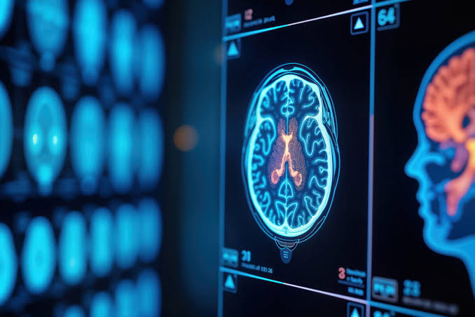
Neuroimaging refers to medical imaging tests that create pictures of the brain, spine, spinal cord, nerves, and blood vessels. These images give doctors information that cannot always be obtained through a physical examination alone. MRI and CT are two of the most commonly used methods, but the exact test depends on the condition being investigated. MRI is particularly useful when detailed images of brain or spinal tissues are needed, while CT can provide rapid information in situations such as head injury or suspected bleeding. Specialised imaging can also show blood vessels or help map important brain functions before selected procedures.
Neuroimaging may help doctors investigate:
• Brain tumours
• Spinal cord conditions
• Head injuries
• Seizure-related abnormalities
• Hydrocephalus
• Stroke and bleeding
• Nerve compression
• Vascular abnormalities
• Selected skull base conditions
Why Is Neuroimaging Important?
Neurological symptoms can have several possible causes. A person with weakness, for example, may have a problem involving the brain, spinal cord, nerve roots, or peripheral nerves. Imaging can help doctors determine whether there is a structural change that may explain the symptoms. It can also show the location and extent of an abnormality and its relationship with nearby structures. This information becomes especially useful when a patient needs treatment or surgery. However, a scan should not be considered a diagnosis by itself. Dr. Ritesh Nawkhare reviews imaging findings together with the patient’s symptoms, neurological examination, medical history, and previous reports. This helps avoid unnecessary treatment and keeps the focus on the actual cause of the patient’s problem.
MRI Brain In Nagpur
Brain MRI provides detailed images of brain tissue and surrounding structures. It does not use ionising radiation and can show differences between various types of soft tissue. Doctors may recommend a brain MRI when they need a closer look at a suspected tumour, seizure-related abnormality, stroke, structural problem, or other neurological condition. MRI can also help monitor a condition that has already been diagnosed and compare changes across different scans. In selected patients, contrast may be used to highlight certain tissues or abnormalities. Some patients may also need specialised MRI techniques, such as functional MRI, when doctors need information about areas involved in speech, movement, memory, or other important functions before brain surgery.
MRI Spine In Nagpur
Spinal MRI gives detailed images of the spinal cord, discs, nerves, vertebrae, and surrounding soft tissues. It can help doctors understand whether symptoms are related to pressure on the spinal cord or nerve roots. Your doctor may recommend an MRI when you have persistent back or neck symptoms, weakness, numbness, changes in sensation, walking difficulties, or other signs that suggest a spinal problem. MRI may also help evaluate spinal tumours, infections, degenerative changes, and selected congenital conditions. In suspected spinal tumours, MRI can provide detailed information about the spinal cord and surrounding tissues, while CT may add useful information about the bones.
CT Brain Scan In Nagpur
A CT brain scan uses X-rays and computer processing to create cross-sectional images of the brain and skull. It is particularly useful when doctors need information quickly. For example, CT is often used during the initial assessment of significant head injuries or suspected brain bleeding. It can also show skull fractures, swelling, and other urgent changes. MRI may provide more detailed images of brain tissue, but it usually takes longer and may not be the first choice in an emergency. The decision between CT and MRI depends on the symptoms, urgency, and information the doctor needs. Dr. Ritesh Nawkhare considers these factors before recommending the appropriate imaging study for a patient.
Neuroimaging For Brain Tumours
Imaging is an important part of evaluating a suspected brain tumour. MRI can show where the lesion is located, how large it is, and how it relates to surrounding brain structures. It may also provide information about swelling or changes around the lesion. These details can help the neurosurgeon understand whether the lesion may be suitable for observation, further testing, biopsy, surgery, or another form of treatment. In some cases, specialized imaging can provide additional information about blood flow, tissue characteristics, or important brain pathways. However, imaging does not always identify the exact tumour type. Some patients may need tissue examination to confirm the diagnosis. The treatment plan therefore depends on the scan, symptoms, examination, and overall clinical picture.
Neuroimaging For Head Injuries
A head injury can affect the brain, skull, or blood vessels, even when there is little or no visible injury on the outside. Imaging helps doctors look for problems such as bleeding, swelling, fractures, or other structural changes. CT is often useful when a rapid assessment is needed because it can quickly show several important traumatic changes. Depending on the injury and the patient’s symptoms, doctors may recommend further imaging, including MRI. A patient should receive urgent medical attention after significant trauma if they develop loss of consciousness, repeated vomiting, worsening headache, seizures, confusion, unusual drowsiness, weakness, or difficulty speaking. The choice of imaging depends on the severity of the injury and the findings during the neurological assessment.
Neuroimaging For Spine And Spinal Cord Problems
Problems affecting the spine can cause pain, weakness, numbness, altered sensation, or difficulty walking. When symptoms suggest pressure or disease involving the spinal cord or nerve roots, imaging can help identify the structural cause. MRI is particularly useful because it provides detailed images of the spinal cord, nerves, discs, and surrounding soft tissues. CT may also be useful when doctors need more information about bone structures or spinal stability. The scan is interpreted together with the patient’s symptoms and neurological examination. This is important because an abnormal scan does not automatically mean that surgery is necessary. Some findings may only need observation, medication, physiotherapy, or regular follow-up. Treatment depends on the underlying problem and how it affects the individual patient.
Neuroimaging And Minimally Invasive Neurosurgery
Minimally invasive neurosurgery requires careful planning. When the surgeon works through a smaller or more targeted route, detailed knowledge of the anatomy becomes even more important. Preoperative imaging can help identify the location of a lesion and show the structures surrounding it. This information may help the surgeon decide whether a minimally invasive approach is appropriate.
However, minimally invasive surgery is not automatically the right choice for every patient. A larger or different surgical approach may sometimes provide better access or control depending on the condition. The decision depends on the lesion, its location, its relationship with important structures, and the overall treatment goal. At Neurosys Multispeciality Centre In Nagpur, Dr. Ritesh Nawkhare considers these factors before discussing the available surgical options.
Neuroimaging For Follow-Up
Neuroimaging can remain useful even after a neurological condition has been diagnosed or treated. Follow-up scans allow doctors to compare current images with previous ones and look for changes over time. Depending on the condition, repeat imaging may show whether a lesion has remained stable, increased in size, or changed after treatment. Follow-up imaging may be considered after brain tumour treatment, spine surgery, head injury, hydrocephalus treatment, vascular procedures, or selected epilepsy interventions. The timing of repeat scans varies from one patient to another. Some conditions need regular monitoring, while others may require imaging only when new symptoms develop. Your doctor decides the follow-up schedule based on your diagnosis, treatment, and clinical progress.
Benefits Of Neuroimaging
Neuroimaging gives doctors a clearer view of structures that cannot be examined directly during a routine consultation. It can help identify abnormalities, clarify the location of a lesion, and provide information that supports diagnosis and treatment planning. MRI can provide detailed images of the brain and spinal cord, while CT can offer rapid assessment in many urgent situations.
The potential benefits include:
• Identifying structural abnormalities
• Supporting diagnosis
• Locating brain or spinal lesions
• Assessing the spinal cord and nerve structures
• Examining selected blood vessel conditions
• Supporting surgical planning
• Monitoring known conditions
• Comparing changes over time
• Helping guide further investigations
The usefulness of imaging depends on the condition being investigated and the specific scan performed.
Why Choose Dr. Ritesh Nawkhare For Neuroimaging Evaluation?
Dr. Ritesh Nawkhare is a Neurosurgeon with MBBS, MS (General Surgery), and MCh (Neurosurgery). His evaluation focuses on understanding the neurological problem as a whole rather than treating an imaging report as a diagnosis on its own. During a consultation, he can review your symptoms, neurological examination, previous medical treatment, and available MRI or CT scans. This can help determine whether an imaging finding is relevant to your symptoms and whether you need further evaluation or treatment. At Neurosys Multispeciality Centre In Nagpur, patients can discuss abnormal brain or spine scans, newly diagnosed lesions, persistent neurological symptoms, or recommendations for neurosurgery. The treatment plan depends on the individual diagnosis, imaging findings, overall health, and clinical needs.
Frequently Asked Questions
Book a Consultation for Neuroimaging in Nagpur
If you have an abnormal brain or spine scan, persistent neurological symptoms, or questions about a recommended neurosurgical procedure, consider scheduling a consultation with Dr. Ritesh Nawkhare. Bring your previous MRI or CT images, reports, prescriptions, and relevant medical records. Reviewing these details together can help clarify the diagnosis and determine whether you need further testing, monitoring, or treatment.
At Neurosys Multispeciality Centre In Nagpur, Dr. Ritesh Nawkhare provides individualized neurosurgical evaluation based on your symptoms, examination, imaging findings, and overall condition.