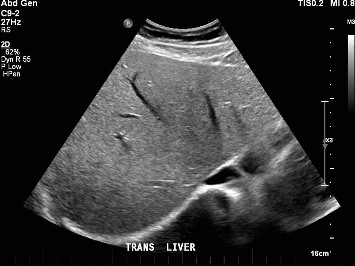
Liver ultrasound
How a Liver Ultrasound Works
During a liver ultrasound, a trained sonographer or radiologist performs the procedure. The sonographer applies a special gel to the patient’s abdomen and uses a handheld device called a transducer. The transducer emits sound waves that travel through the skin and bounce off the liver and surrounding tissues. These sound waves create echoes, which the transducer captures and converts into detailed images displayed on a monitor.
The entire process is painless and typically takes 20 to 30 minutes. Patients do not need any special preparation, although fasting for a few hours before the procedure may be required to ensure clear images.
Benefits of a Liver Ultrasound
A liver ultrasound offers numerous advantages, making it a preferred diagnostic tool for liver-related conditions. It is safe, radiation-free, and does not involve any incisions or injections. The procedure provides real-time imaging, allowing healthcare providers to assess blood flow, detect tumors, and identify structural abnormalities. Additionally, it is cost-effective and widely available in most medical facilities.
Conditions Diagnosed with a Liver Ultrasound
- Liver Cirrhosis: The ultrasound detects changes in liver texture and size, which are common in cirrhosis, a condition caused by chronic liver damage.
- Fatty Liver Disease: The imaging technique identifies fat accumulation in the liver, a hallmark of fatty liver disease.
- Liver Tumors or Cysts: The procedure distinguishes between benign cysts and malignant tumors, aiding in early cancer detection.
- Hepatitis: Inflammation of the liver caused by hepatitis can be assessed through changes in liver appearance on the ultrasound.
- Gallstones or Biliary Obstruction: The ultrasound visualizes the gallbladder and bile ducts, helping identify gallstones or blockages.
- Liver Abscesses or Infections: The imaging technique detects pockets of infection or fluid accumulation in the liver.
- Portal Hypertension: The ultrasound evaluates blood flow in the portal vein, which can indicate increased pressure due to liver disease.
Why Choose a Liver Ultrasound?
A liver ultrasound is a reliable and efficient tool for evaluating liver health. It provides detailed insights without the risks associated with other imaging methods, such as CT scans or MRIs, which involve radiation or contrast agents. The procedure is suitable for patients of all ages, including pregnant women, and can be repeated as needed to monitor liver conditions over time.
Preparing for a Liver Ultrasound
To ensure accurate results, patients should follow specific guidelines before the procedure. These may include fasting for 8 to 12 hours to reduce gas in the intestines, which can obstruct the view of the liver. Patients should also inform their healthcare provider about any medications or medical conditions that might affect the test.
What to Expect During the Procedure
During the liver ultrasound, the patient lies on an examination table, and the sonographer applies a water-based gel to the abdomen. The gel ensures proper contact between the skin and the transducer, allowing sound waves to travel effectively. The sonographer moves the transducer across the abdomen, capturing images from different angles. The patient may be asked to hold their breath briefly to obtain clearer images.
After the Liver Ultrasound
Once the procedure is complete, the sonographer wipes off the gel, and the patient can resume normal activities immediately. A radiologist analyzes the images and sends a report to the referring physician, who discusses the results with the patient and recommends further steps if necessary.Contact Us
Conclusion
A liver ultrasound is a vital diagnostic tool that provides detailed insights into liver health. It is safe, effective, and widely used to detect and monitor various liver conditions. If you experience symptoms such as abdominal pain, jaundice, or unexplained weight loss, consult your healthcare provider to determine if a liver ultrasound is necessary. Early detection and diagnosis can significantly improve treatment outcomes and overall liver health.Schedule your Consultation with Dr. Ritesh Nawkhare