
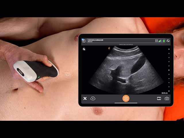
Gallbladder Ultrasound In Nagpur For Detecting Gallstones And Gallbladder Changes with Dr.Ritesh Nawkhare
Gallbladder Ultrasound In Nagpur is a non-invasive diagnostic examination that uses high-frequency sound waves to create detailed images of the gallbladder, bile ducts, liver, and nearby abdominal structures. It is commonly recommended when symptoms such as upper abdominal pain, nausea, vomiting, indigestion, bloating, or jaundice may indicate a gallbladder or biliary concern. The examination can help identify gallstones, biliary sludge, gallbladder wall thickening, enlargement, fluid around the gallbladder, and changes in the visible bile ducts. A gallbladder ultrasound can also provide valuable information when liver-related blood tests are abnormal or when a previously identified gallbladder condition needs follow-up. Because the examination does not use ionising radiation, it is widely used as an initial imaging method for many abdominal concerns. At NNS Hospital, Gallbladder Ultrasound In Nagpur can form part of a structured diagnostic evaluation. The ultrasound findings can be reviewed alongside your symptoms, medical history, physical examination, laboratory results, and previous imaging to help your healthcare provider understand the underlying concern and decide whether further testing or specialist evaluation may be appropriate.
Understanding Gallbladder Ultrasound
The gallbladder is a small organ located beneath the liver. It stores and concentrates bile, which helps the body digest dietary fats. Changes affecting the gallbladder can sometimes cause noticeable symptoms, while other findings may be discovered during an examination for another abdominal concern.
During an ultrasound, a transducer sends high-frequency sound waves through the upper abdomen. The returning echoes are converted into images that allow the sonographer to examine the gallbladder and related structures.
The assessment may include:
▪ Gallbladder size and shape
▪ Gallbladder wall appearance
▪ Internal contents
▪ Presence of stones or sludge
▪ Bile duct appearance
▪ Surrounding abdominal structures.
When Is Gallbladder Ultrasound Recommended?
Your doctor may recommend an ultrasound when symptoms or clinical findings suggest a possible gallbladder or biliary problem. The scan can also be useful for monitoring a previously identified abnormality.
Common reasons for the examination include:
▪ Pain in the upper right or upper central abdomen
▪ Recurrent abdominal discomfort after meals
▪ Nausea or vomiting
▪ Suspected gallstones
▪ Possible gallbladder inflammation
▪ Jaundice or suspected bile flow problems
▪ Abnormal liver function tests
▪ Unexplained digestive symptoms
▪ Follow-up of a known gallbladder condition
The need for imaging depends on your individual symptoms, medical history, and clinical examination.
What Can A Gallbladder Ultrasound Show?
Ultrasound provides real-time images of the gallbladder and can reveal several structural changes. It is particularly useful for identifying many gallstones and assessing the gallbladder wall and surrounding anatomy.
The examination may help identify:
▪ Gallstones
▪ Biliary sludge
▪ Gallbladder wall thickening
▪ Gallbladder distension
▪ Polyps or focal abnormalities
▪ Fluid around the gallbladder
▪ Enlargement of the bile ducts
▪ Other structural changes requiring further assessment
An ultrasound finding does not always establish the final diagnosis. Your healthcare provider may correlate the results with symptoms, blood tests, and other investigations.
Gallbladder Ultrasound For Gallstone Detection
Gallstones are hardened deposits that can form inside the gallbladder. Some people have no symptoms, while others may experience episodes of abdominal pain, particularly after eating. Ultrasound is commonly used to look for gallstones and related gallbladder changes. During the examination, the sonographer may assess the size, location, and appearance of visible stones along with the condition of the gallbladder.
Depending on the findings, your doctor may consider:
▪ Frequency and severity of symptoms
▪ Number and appearance of stones
▪ Gallbladder wall changes
▪ Bile duct findings
▪ Associated blood test abnormalities
Further treatment or specialist consultation depends on the complete clinical picture.
Assessing Gallbladder Inflammation
Inflammation of the gallbladder can develop for different reasons and may cause significant abdominal symptoms. Ultrasound can provide important structural information when inflammation is suspected.
The examination may assess:
▪ Gallbladder wall thickness
▪ Gallbladder distension
▪ Internal contents
▪ Fluid surrounding the gallbladder
▪ Tenderness during probe pressure when clinically assessed
▪ Associated biliary changes
These findings are interpreted together with symptoms, physical examination, and laboratory results rather than in isolation.
Bile Duct Evaluation During Ultrasound
The bile ducts carry bile from the liver and gallbladder toward the digestive tract. A blockage or other biliary change can sometimes affect bile flow and may be associated with jaundice or abnormal liver tests. Ultrasound can assess the visible portions of the bile ducts and may identify enlargement that requires further evaluation.
Possible reasons for additional assessment include:
▪ Suspected bile duct obstruction
▪ Jaundice
▪ Abnormal liver function tests
▪ Gallstones with concerning symptoms
▪ Changes identified during an earlier examination
If the ultrasound suggests a biliary problem, your doctor may recommend additional imaging or laboratory investigations.
Gallbladder Ultrasound For Abdominal Pain
Upper abdominal pain can have several possible causes. The gallbladder is one of the structures that may need to be evaluated when symptoms occur in the upper right or central abdomen.
A gallbladder ultrasound may be considered when pain is:
▪ Recurrent or persistent
▪ Associated with meals
▪ Accompanied by nausea or vomiting
▪ Associated with jaundice
▪ Associated with abnormal liver-related tests
Ultrasound can provide useful anatomical information, but your healthcare provider will consider other possible causes of abdominal pain as well.
Preparation Before Gallbladder Ultrasound
Proper preparation can help improve the quality of abdominal ultrasound images. Your healthcare provider may ask you to avoid food and drinks for a specified period before the examination.
Before your appointment:
▪ Follow the fasting instructions provided by the imaging team
▪ Take medicines according to the instructions given to you
▪ Inform the healthcare team about relevant medical conditions
▪ Bring previous ultrasound or abdominal imaging reports if available
▪ Follow any additional instructions provided before the scan
If fasting is difficult because of a medical condition or medication requirement, discuss this with your healthcare provider beforehand.
What Happens During Gallbladder Ultrasound?
Gallbladder ultrasound is generally performed as an outpatient examination. You will usually lie comfortably on an examination table while the sonographer positions you for appropriate imaging.
A small amount of gel is placed on the skin of the upper abdomen. The transducer is then moved gently across the area to obtain images from different angles.
You may be asked to:
▪ Lie on your back or side
▪ Take a deeper breath when requested
▪ Hold your breath briefly
▪ Remain still during image capture
The procedure does not normally involve an incision, injection, or anaesthesia.
Benefits Of Gallbladder Ultrasound In Nagpur
Gallbladder ultrasound provides a practical way to examine the gallbladder and biliary system when symptoms or clinical findings require imaging.
Key benefits include:
▪ Non-invasive imaging
▪ No ionising radiation
▪ Useful for detecting many gallstones
▪ Assessment of gallbladder wall and internal contents
▪ Evaluation of visible bile ducts
▪ Real-time imaging during the examination
▪ Can support investigation of upper abdominal symptoms
▪ Useful for follow-up of selected gallbladder findings
▪ Usually requires no recovery time
▪ Can help guide decisions about additional investigations.
Recovery After Gallbladder Ultrasound
Most people can return to their normal activities immediately after a routine gallbladder ultrasound. There is generally no recovery period because the examination does not involve surgery or anaesthesia.
If you were asked to fast before the scan, you can usually resume eating and drinking according to the instructions provided after the examination.
▪ No surgical wound is created
▪ No routine recovery period is required
▪ Normal activities can generally continue
▪ Follow-up depends on the examination findings
▪ Additional testing may be recommended when necessary
Your doctor can explain the report and discuss whether any further evaluation is appropriate.
Why Choose NNS Hospital For Gallbladder Ultrasound?
At NNS Hospital, gallbladder ultrasound can form part of a coordinated diagnostic assessment based on your symptoms and medical requirements. The findings can be considered alongside clinical history, laboratory investigations, and other imaging when needed.
The approach focuses on:
▪ Appropriate imaging based on the clinical indication
▪ Careful assessment of the gallbladder and biliary structures
▪ Clear preparation guidance
▪ Patient-friendly communication
▪ Correlation of imaging findings with clinical information
▪ Appropriate recommendations for further evaluation when required
The multispeciality setting can support coordination between different healthcare services when a patient’s condition requires broader evaluation.
Frequently Asked Questions
Book a Consultation for Gallbladder ultrasound in Nagpur
If you have recurring upper abdominal discomfort, suspected gallstones, digestive symptoms, jaundice, or abnormal liver-related test results, your doctor may recommend gallbladder imaging as part of your evaluation. At NNS Hospital, the examination can be planned according to your clinical needs. Bring previous reports and relevant investigation results to help your healthcare provider understand your medical history and determine the appropriate diagnostic pathway.
Book a consultation for Gallbladder Ultrasound In Nagpur and take a timely step toward understanding your abdominal and biliary health.