
Thyroid Ultrasound In Nagpur For Advanced Evaluation Of Thyroid And Neck Conditions with Dr.Ritesh Nawkhare
Thyroid Ultrasound In Nagpur is a detailed, non-invasive imaging examination that helps evaluate the thyroid gland and selected structures in the neck. It can provide important information when a person has a thyroid swelling, neck lump, palpable nodule, abnormal thyroid examination, or a previously detected thyroid lesion that requires follow-up. Unlike blood tests, which mainly assess thyroid function, ultrasound provides structural information. It can show the size and shape of the thyroid, identify nodules, assess their internal appearance, and examine selected surrounding tissues. This information can help your healthcare professional decide whether observation, repeat imaging, laboratory testing, or further investigation may be appropriate. At Neurosys, thyroid imaging is approached as part of a broader clinical evaluation. Symptoms, examination findings, previous reports, thyroid function tests, medical history, and the ultrasound appearance are considered together. This helps create a more meaningful assessment instead of interpreting an imaging finding in isolation.
What Does A Thyroid Ultrasound Assess?
The thyroid is a small gland located at the front of the neck, but even subtle structural changes can sometimes require detailed assessment. Ultrasound provides real-time images of the gland and can document changes that may not be obvious through physical examination alone.
A thyroid ultrasound may assess:
▪ Overall thyroid size and shape
▪ Right and left thyroid lobes
▪ Thyroid nodules and their measurements
▪ Solid, cystic, or mixed lesions
▪ Internal characteristics of visible nodules
▪ Nodule margins and shape
▪ Selected cervical lymph nodes
▪ Changes compared with previous imaging
The findings can help determine whether a thyroid abnormality requires observation or additional clinical evaluation.
When Is Thyroid Ultrasound Recommended?
Your healthcare professional may recommend thyroid ultrasound when there is a specific clinical reason to examine the gland or neck. It can be particularly useful when a structural abnormality has been detected or suspected.
Common situations include:
▪ A newly noticed lump in the neck
▪ Thyroid swelling or enlargement
▪ A nodule felt during examination
▪ Follow-up of a known thyroid nodule
▪ Changes in an existing thyroid lesion
▪ An incidental thyroid finding on another scan
▪ Selected cervical lymph node concerns
The need for ultrasound depends on your symptoms and clinical findings. It is not necessary for every person with a thyroid-related concern.
Detailed Assessment Of Thyroid Nodules
Thyroid nodules can vary considerably in appearance. Some contain fluid, some are predominantly solid, and others have a combination of features. Ultrasound helps document these characteristics in greater detail.
The examination may evaluate:
▪ Nodule size and location
▪ Composition and internal structure
▪ Shape and orientation
▪ Margins
▪ Internal echogenicity
▪ Selected calcification patterns
▪ Associated neck findings
These features can contribute to risk assessment and help determine whether a nodule can be monitored or needs additional evaluation.
Importantly, finding a thyroid nodule does not automatically mean that it is cancerous. Many thyroid nodules are benign. Clinical history and imaging characteristics help guide the next step.
Thyroid Ultrasound For Cysts And Complex Lesions
Not every thyroid lump represents the same type of abnormality. Some lesions are predominantly fluid-filled, while others contain both fluid and solid components. Ultrasound can help distinguish between these appearances and document the size and internal characteristics of the lesion.
This information may help your healthcare professional decide whether:
▪ Observation is appropriate
▪ Repeat ultrasound is useful
▪ Laboratory assessment is needed
▪ Specialist evaluation should be considered
▪ Tissue sampling may be appropriate
The management decision depends on the complete clinical assessment.
Ultrasound-Guided Evaluation Of Thyroid Nodules
When a thyroid nodule requires tissue sampling, ultrasound can provide important guidance for selecting and targeting the appropriate area. A fine-needle aspiration may be recommended for selected nodules based on factors such as their ultrasound appearance, size, clinical history, and overall risk assessment.
Ultrasound guidance can help the healthcare professional visualise the lesion during the procedure and obtain a sample from the intended area.
Not every thyroid nodule requires aspiration. In many cases, careful monitoring may be appropriate.
Thyroid Ultrasound And Thyroid Function Tests
Thyroid ultrasound and thyroid blood tests provide different types of information.
Blood tests can help determine whether the thyroid is producing an appropriate amount of thyroid hormone. Ultrasound, in contrast, evaluates the physical structure of the gland.
Depending on your symptoms and clinical findings, your evaluation may include:
▪ Thyroid-stimulating hormone assessment
▪ Other thyroid hormone tests when indicated
▪ Physical examination
▪ Thyroid ultrasound
▪ Review of previous imaging
▪ Additional testing when clinically required
A person can have a structural thyroid nodule even when thyroid hormone levels are normal. Therefore, test results need to be interpreted together.
Thyroid And Neck Lymph Node Assessment
A thyroid ultrasound may also provide information about selected lymph nodes in the surrounding neck region.
When a thyroid abnormality is being assessed, examining relevant neck structures can add useful context to the imaging evaluation.
The report may describe lymph node size and selected structural characteristics when these are visible and clinically relevant.
If a finding requires further assessment, your healthcare professional can explain whether additional imaging, monitoring, or tissue evaluation is appropriate.
How Is Thyroid Ultrasound Performed?
A thyroid ultrasound is usually a straightforward outpatient examination.
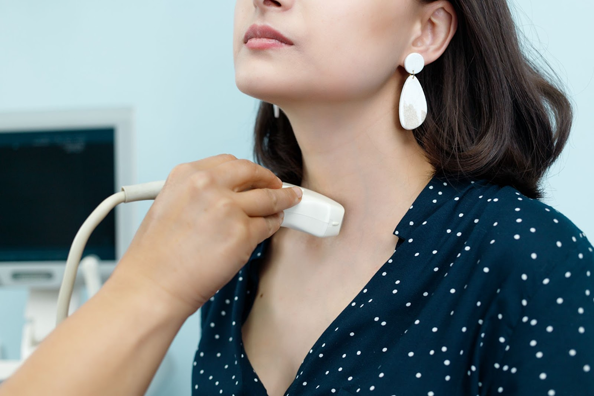
You lie comfortably on your back while the neck is positioned to provide access to the thyroid region. A small amount of gel is applied to the skin, and the ultrasound transducer is gently moved across the front and sides of the neck.
The healthcare professional captures images from different angles to assess the thyroid and relevant surrounding structures.
▪ No incision is required
▪ No injection is normally needed
▪ No ionising radiation is used
▪ The examination is generally painless
▪ Images are obtained in real time
▪ Normal activities can usually continue afterwards.
How Should You Prepare For Thyroid Ultrasound?
Most standard thyroid ultrasound examinations do not require extensive preparation.
Before your appointment, it can be useful to:
▪ Wear clothing that allows comfortable access to the neck
▪ Bring previous ultrasound reports
▪ Carry relevant thyroid blood test results
▪ Inform the healthcare professional about previous thyroid procedures
▪ Mention any newly noticed changes in the neck
Previous reports can be particularly useful when the examination is being performed to monitor an existing thyroid nodule.
When Is Follow-Up Imaging Needed?
Some thyroid abnormalities can be safely monitored over time rather than treated immediately. Follow-up ultrasound may be suggested when your healthcare professional wants to assess whether a known lesion remains stable.
During follow-up, imaging may compare:
▪ Nodule measurements
▪ Internal appearance
▪ Margins and shape
▪ Development of new features
▪ Surrounding neck findings
The timing of repeat imaging depends on the individual finding and clinical assessment. It is not the same for every thyroid nodule.
Recovery After Thyroid Ultrasound
A routine thyroid ultrasound does not usually require a recovery period. Since the examination is non-invasive and does not normally involve an injection or incision, most people can return to their regular activities immediately.
▪ Resume normal activities after the scan
▪ Review the report with your healthcare professional
▪ Keep previous reports for future comparison
▪ Attend follow-up appointments when advised
▪ Complete additional investigations if recommended
If a separate procedure, such as a biopsy, is performed, its aftercare may be different.
Why Choose Neurosys For Thyroid Ultrasound In Nagpur?
A thyroid ultrasound is most useful when its findings are understood in the context of the patient’s overall health. At Neurosys, the focus is on structured imaging, clear communication, and appropriate next-step planning.
▪ Detailed assessment of the thyroid gland
▪ Evaluation of thyroid nodules and structural changes
▪ Review of previous imaging when available
▪ Consideration of relevant clinical information
▪ Appropriate guidance for further investigation
▪ Clear explanation of imaging findings
▪ Individualised follow-up when required
The goal is to provide useful diagnostic information while avoiding unnecessary testing or assumptions based on a single finding.
Frequently Asked Questions
It is a non-invasive imaging examination that uses sound waves to assess the thyroid gland, thyroid nodules, and selected surrounding neck structures.
Book a Consultation for Thyroid Ultrasound in Nagpur
A neck lump, thyroid swelling, or previously detected thyroid nodule deserves appropriate evaluation. A consultation can help determine whether ultrasound is suitable and what additional assessment may be needed.
During your consultation, you may discuss:
▪ Your current symptoms or neck concerns
▪ Previous thyroid investigations
▪ Relevant blood test results
▪ Existing thyroid nodules or swelling
▪ The need for imaging or follow-up
▪ Possible next steps after the ultrasound
Book a consultation for Thyroid Ultrasound In Nagpur at Neurosys for a detailed and personalized thyroid assessment.