Fetal echocardiography
Fetal echocardiography is a specialized ultrasound examination that evaluates the structure and function of a baby’s heart while still in the womb. This advanced diagnostic tool plays a critical role in detecting congenital heart defects (CHDs) early, allowing healthcare providers to plan appropriate care before and after birth. In this detailed guide, we will explore the importance of fetal echocardiography, how it works, its benefits, and what to expect during the procedure.
What is Fetal Echocardiography?
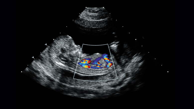
Fetal echocardiography is a non-invasive imaging technique that uses high-frequency sound waves to create detailed images of the fetal heart. Unlike a standard prenatal ultrasound, which provides a general overview of the baby’s development, fetal echocardiography focuses specifically on the heart’s anatomy, blood flow, and rhythm.
Fetal echocardiography is a non-invasive imaging technique that uses high-frequency sound waves to create detailed images of the fetal heart. Unlike a standard prenatal ultrasound, which provides a general overview of the baby’s development, fetal echocardiography focuses specifically on the heart’s anatomy, blood flow, and rhythm.
Why is Fetal Echocardiography Important?
Congenital heart defects are among the most common birth defects, affecting nearly 1% of all live births worldwide. Early detection of these conditions is crucial for several reasons:
- Early Intervention: Identifying heart defects before birth allows healthcare providers to plan for specialized care immediately after delivery.
- Improved Outcomes: Early diagnosis often leads to better management and treatment options, improving the baby’s overall prognosis.
- Parental Preparedness: Parents can prepare emotionally and logistically for the birth of a child with a heart condition, including choosing a hospital with the necessary resources.
- Pregnancy Management: In some cases, fetal echocardiography may influence decisions about the timing and mode of delivery.
Who Should Undergo Fetal Echocardiography?
While fetal echocardiography is not routinely performed on all pregnant women, it is recommended in specific high-risk situations, including:
- Family History: A family history of congenital heart defects increases the likelihood of the baby having a similar condition.
- Maternal Health Conditions: Conditions such as diabetes, lupus, or phenylketonuria (PKU) can elevate the risk of fetal heart defects.
- Medication Exposure: Certain medications taken during pregnancy, such as anti-seizure drugs, may increase the risk of CHDs.
- Abnormal Routine Ultrasound: If a standard prenatal ultrasound reveals potential heart abnormalities, fetal echocardiography is often recommended.
- Multiple Pregnancies: Twins or higher-order multiples have a higher risk of congenital heart defects.
- Advanced Maternal Age: Women over 35 years old are at a slightly higher risk of having a baby with a heart defect.
- Previous Pregnancy Complications: A history of miscarriages or stillbirths may warrant closer monitoring.
How Does Fetal Echocardiography Work?
- Preparation: The procedure requires no special preparation. The pregnant woman lies comfortably on an examination table, and a water-based gel is applied to her abdomen to improve sound wave transmission.
- Transducer Placement: A handheld device called a transducer is moved over the abdomen. The transducer emits sound waves that bounce off the baby’s heart and create echoes.
- Image Creation: The echoes are converted into real-time images of the heart, which are displayed on a monitor.
- Analysis: A trained specialist, usually a pediatric cardiologist or maternal-fetal medicine specialist, analyzes the images to assess the heart’s structure, function, and blood flow.
Types of Fetal Echocardiography
- Transabdominal Echocardiography: This is the most common approach, where the transducer is placed on the mother’s abdomen.
- Transvaginal Echocardiography: In some cases, especially during early pregnancy, a transvaginal approach may be used for better image clarity.
What Can Fetal Echocardiography Detect?
- Septal Defects: Holes in the walls separating the heart’s chambers (e.g., atrial septal defect or ventricular septal defect).
- Valve Abnormalities: Issues with the heart valves, such as stenosis or atresia.
- Hypoplastic Left Heart Syndrome: Septal Defects: Holes in the walls separating the heart’s chambers (e.g., atrial septal defect or ventricular septal defect).
- Valve Abnormalities: Issues with the heart valves, such as stenosis or atresia.
- Hypoplastic Left Heart Syndrome: Underdevelopment of the left side of the heart.
- Tetralogy of Fallot: A combination of four heart defects affecting blood flow.
- Transposition of the Great Arteries: A condition where the two main arteries leaving the heart are switched.
- Arrhythmias: Irregular heart rhythms that may require treatment.
Benefits of Fetal Echocardiography
- Early Diagnosis: Detecting heart defects before birth allows for timely intervention.
- Accurate Assessment: Provides detailed information about the heart’s structure and function.
- Guided Treatment Plans: Helps healthcare providers develop a tailored care plan for the baby.
- Peace of Mind: Reassures parents when no abnormalities are found.
Limitations of Fetal Echocardiography
- Operator Dependency: The accuracy of the results depends on the skill and experience of the technician.
- False Positives/Negatives: In rare cases, the procedure may miss a defect or suggest a problem that doesn’t exist.
- Late-Onset Defects: Some heart conditions develop later in pregnancy or after birth and may not be detected during the scan.
What to Expect After the Procedure
- After the fetal echocardiography, the specialist will discuss the findings with the parents. If a heart defect is detected, the healthcare team will explain the condition, its implications, and the next steps. This may include additional tests, consultations with pediatric cardiologists, or planning for delivery at a specialized center.
The Role of Advanced Technology in Fetal Echocardiography
- Advancements in ultrasound technology have significantly improved the accuracy and capabilities of fetal echocardiography. Techniques such as 3D/4D imaging, Doppler ultrasound, and speckle tracking echocardiography provide even more detailed insights into the fetal heart’s structure and function.Contact Us
Conclusion
Fetal echocardiography is a vital tool in modern prenatal care, offering early detection and management of congenital heart defects. By identifying potential issues before birth, healthcare providers can ensure the best possible outcomes for both mother and baby. If you are at risk or have concerns about your baby’s heart health, consult your healthcare provider to determine if fetal echocardiography is right for you.Schedule your Consultation with Dr. Ritesh Nawkhare